
Figure 9.
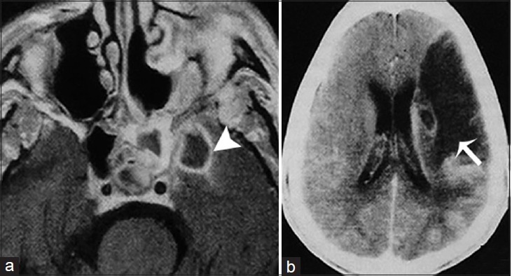
A 51-year-old man, who presented with right-sided paresis and had a history of uncontrolled diabetes mellitus, is diagnosed with chronic invasive fungal Mucormycosis. (a) Magnetic resonance imaging T1 postcontrast imaging of the brain in axial view demonstrates an infectious process in the left sphenoidal sinus and an intracranial intraparenchymal ring enhancing extension in the medial temporal lobe indicative of an abscess (arrowhead). (b) Contrast-enhanced computer tomography image of the same patient shows and acute infarct in the vascular territory of the left middle cerebral artery secondary to intracranial vasculitis (arrow).