
Figure 3.
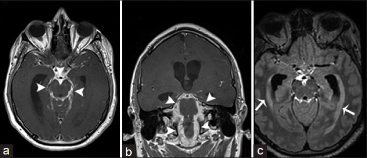
A 25-year-old man, who presented with a history of acute daily headaches over the past 6 months and had a history of chronic coccidioidal meningitis (treated), is diagnosed with chronic hypertrophic basal arachnoiditis and secondary hydrocephalus. Magnetic resonance imaging T1-weighted postcontrast imaging of the brain in axial (a) and coronal (b) view demonstrates marked leptomeningeal hypertrophy with diffuse enhancement involving predominantly the basal cisterns (arrowheads). (c) Magnetic resonance imaging fluid-attenuated inversion recovery postcontrast imaging of the brain in axial view shows a significant enlargement of the lateral ventricles with transependymal migration of cerebrospinal fluid (arrows). These findings are consistent with a hydrocephalus secondary to chronic hypertrophic basal arachnoiditis.