
Figure 6.
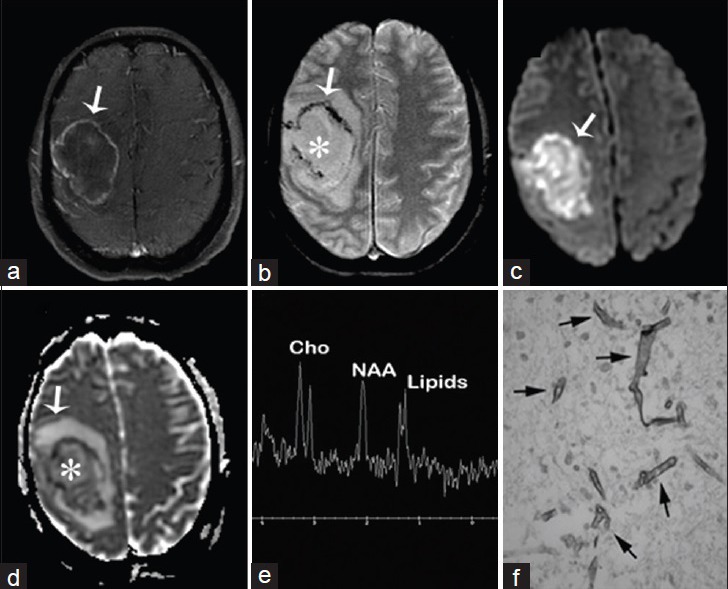
A 50-year-old woman with acute myeloid leukemia, fever, headaches, and neurologic deficits diagnosed with brain abscess due to Rhizomucor pusillus (a) Magnetic resonance imaging T1 fast spin echo/fat sat postcontrast of the brain, axial view shows right sided mass with enhancing wall (arrow) (b) magnetic resonance imaging T2* GRE, axial view shows nonenhancing liquefied core (asterisk) and hypointense wall with susceptibility artifact (arrow) (c and d) diffusion-weighted imaging and apparent diffusion coefficient map of the brain, axial view show restricted diffusion of the wall and intracavitary projections (arrows) and no restriction inside the lesion (asterisk d) (e) MR spectroscopy of the brain shows inverted NAA/Cho ratio due to NAA depletion and increased lipids due to cell destruction (f) Gomori methenamine silver nitrate stain shows Zygomycetes (arrows).