
Figure 2.
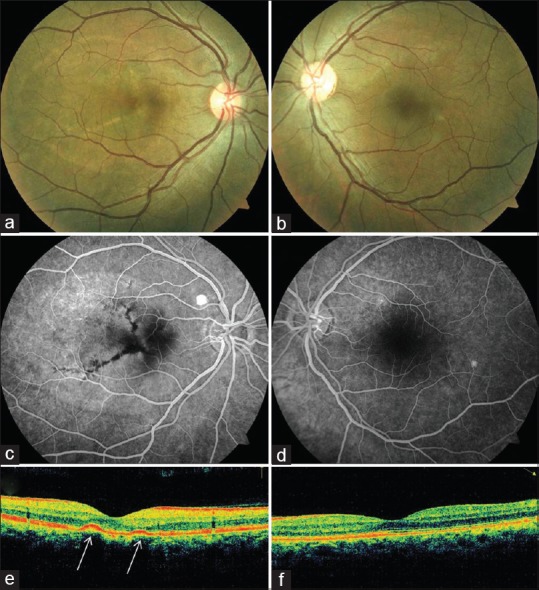
After 3 months, flattening of the pigment epithelial detachments (PEDs) was noted clinically (a and b), angiographically (c and d) and on spectral domain optical coherence tomography (e and f) (except one PED superotemporal to the right disc). In the right eye, linear streaks of altered pigmentation persisted, which blocked fluorescence and were likely retinal pigment epithelium hyperplasia (arrows).