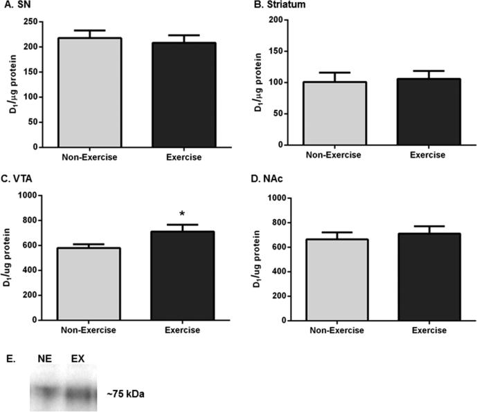
Figure 7.
DA-D1 receptor expression in the nigrostriatal and mesoaccumbens pathways following exercise. Results are expressed as D1 per microgram of protein, and a two-tailed unpaired t-test was used to determine relative differences in DA-D1 receptor expression between the two groups. Data are presented as mean ± SEM. (A) SN. D1 receptor protein expression was not significantly different in exercise rats compared to that in non-exercise rats (n = 8 per group, t = 0.440, p = 0.67). (B) Striatum. D1 receptor expression was not significantly different following exercise (n = 8 (non-exercise), n = 7 (exercise), t = 0.255, p = 0.80). (C) VTA. D1 receptor expression was significantly increased following exercise (n = 8 per group, t = 2.152, *p < 0.05). (D) NAc. D1 receptor protein expression was not significantly different between the groups (n = 8 per group, t = 0.517, p = 0.58). (E) Representative western blot of DA-D1 receptor immunoreactivity. A representative blot showing the effect of exercise upon D1 receptor immunoreactivity in the VTA of one exercise and one non-exercise rat.

