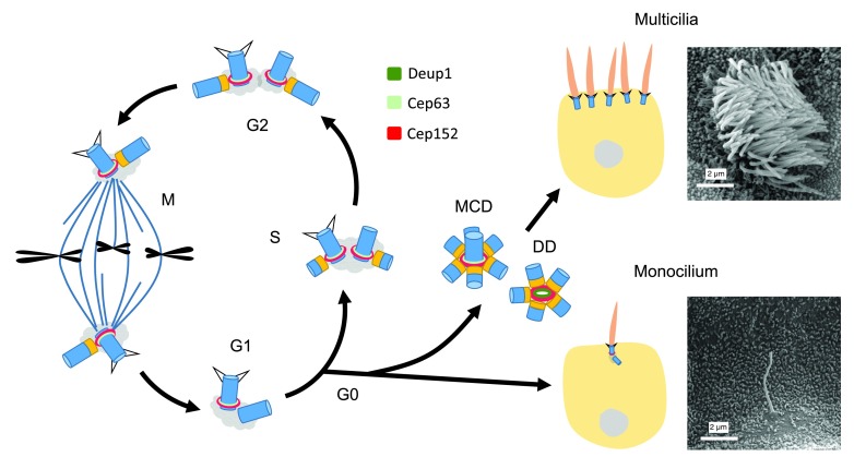
Figure 1. Centriole biogenesis and cilia formation.
The centrosome in a G1 cell contains a pair of mother-daughter centrioles. Upon entering the S phase, each centriole starts to duplicate one daughter centriole so that the centriole number remains constant after mitosis ( a). When the cell enters G0, the mother centriole can be transformed into the basal body to support monocilium formation ( b). Alternatively, both the mother centriole-dependent (MCD) and deuterosome-dependent (DD) pathways can be activated to generate an abundance of centrioles for dense multicilia formation ( c). The scanning electron microscopy images show a primary cilium in the collecting duct of mouse kidney and multicilia of a multiciliated cell in mouse tracheal epithelium, respectively. Centrioles are drawn in blue and their cartwheels in orange.

