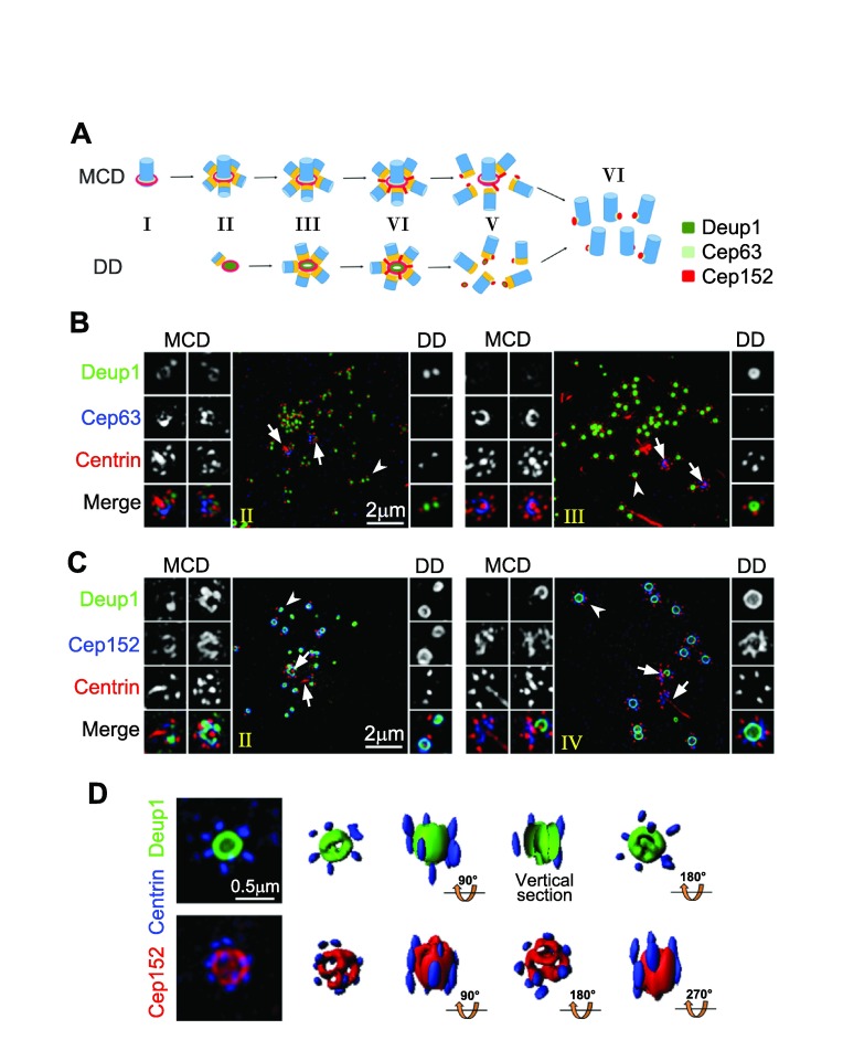
Figure 2. Centriole amplifications in mouse tracheal epithelial cells (MTECs) and mouse ependymal cells (MEPCs).
( A) Illustration for centriole amplification stages in MTECs 19. Centrioles are drawn in blue and their cartwheels in orange. ( B) Three-dimensional structured illumination microscopy (3D-SIM) images for MTECs at early stages (II and III) of centriole amplification. MTECs cultured as described previously 19 were immunostained for Deup1, Cep63, and Centrin and imaged using a DeltaVision OMX V3 microscopic system (GE Healthcare). The mother centrioles (arrows) and representative deuterosomes (arrowheads) are magnified 2× to show details. ( C) 3D-SIM images showing centriole amplification in MEPCs. MEPCs were isolated from neonatal C57BL/6J mice and cultured as described 32. The cells were fixed at day three after serum starvation and immunostained for Deup1, Cep152, and Centrin. The stages (II and IV) are defined as in the MTECs. Note that MEPC deuterosomes ( C) are usually much larger than those in MTECs ( B). ( D) SIM images of two large MEPC deuterosomes immunostained for Deup1 and Centrin (top row) or Cep152 and Centrin (bottom row). Their 3D profiles are also shown. Abbreviations: DD, deuterosome dependent; MCD, mother centriole dependent.

