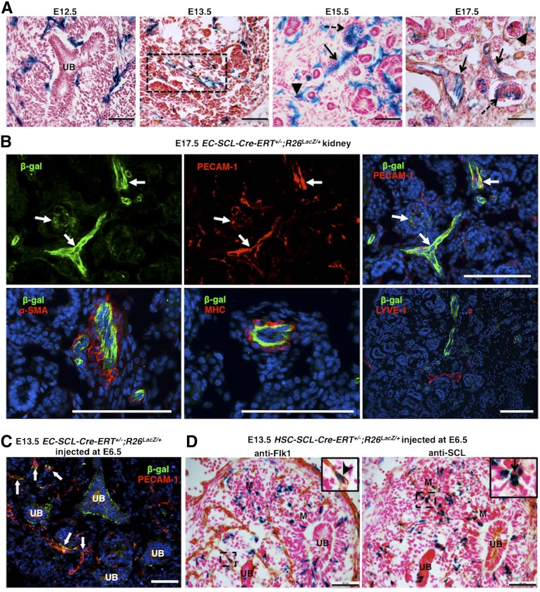
Figure 1.
SCL+ endothelial precursors give rise to renal endothelium but not mural cells in the embryonic kidney. (A) Kidneys of EC-SCl-Cre-ERT+/−;R26LacZ/+ mice treated with tamoxifen at E9.5 show EC-SCL progenitor–derived cells labeled by X-gal staining in blue (β-gal+) distributed within the metanephric mesenchyme surrounding the branching ureteric bud (UB) at E12.5, in a nascent vessel (dashed rectangle) at E13.5 and in ECs of renal arteries and arterioles (arrows), peritubular capillaries (arrowheads), and glomerular capillaries (dashed arrows) at E15.5 and E17.5. (B) β-gal+ (green) cells coexpress the EC marker PECAM-1 (red) (arrows, upper panels), but not the mural markers α-SMA (red) and myosin heavy chain (red), or the lymphatic EC marker LYVE-1 (red) (lower panels). Nuclei were stained with Hoechst (blue). (C) E13.5 EC-SCl-Cre-ERT+/−;R26LacZ/+ mice treated with tamoxifen at E6.5 show several β-gal+ cells (green) costained with PECAM-1 (red) in the developing vessels (arrows) of the kidney. Nuclei were stained with Hoechst (blue). (D) Immunostaining of E13.5 kidney consecutive sections from HSC-SCl-Cre-ERT+/−;R26LacZ/+ mice treated with tamoxifen at E6.5 showed that only few β-gal+ cells coexpressed the early EC marker Flk1(inset, arrow head). Instead, most of them were distributed in the undeveloped mesenchyme (M) and coexpressed SCL/Tal1 (inset, arrow). Scale bars: 50 μm (A, C, and D) and 100 μm (B).

