Figure 6.
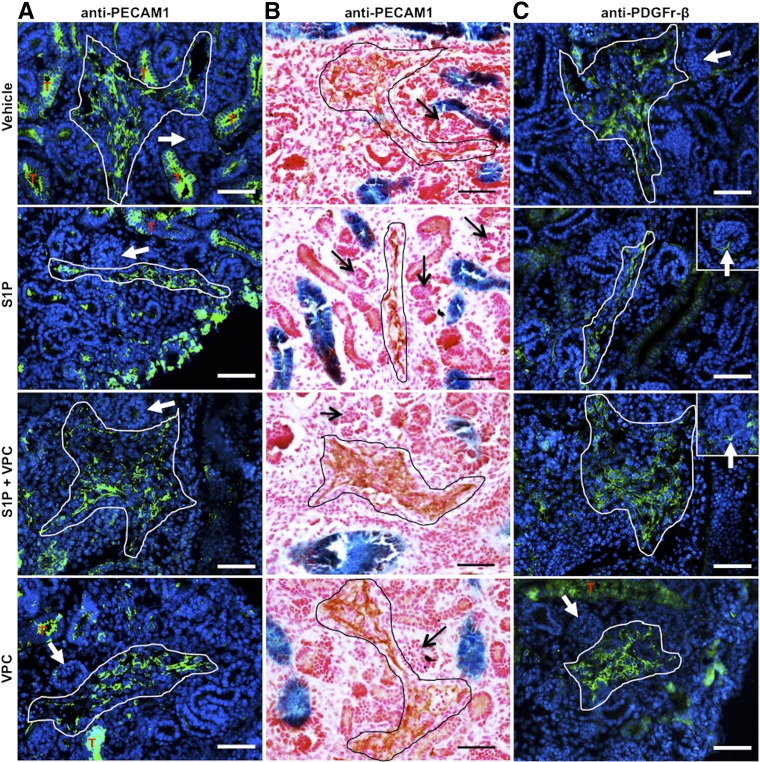
Kidney vascular development ex vivo requires S1P-S1P1 signaling. E12.5 Hoxb7Cre+/−;R26LacZ/+ kidneys treated with vehicle, S1P, S1P+VPC, and VPC in culture for 72 hours and subjected to immunostaining for PECAM-1 and PDGFR-β. (A) Immunofluorescence for PECAM-1 (in green) showed that control kidneys (vehicle) and kidneys treated with VPC developed thickened EC layer (white lined area), whereas those treated with S1P showed a thin layer of ECs. Kidneys treated with S1P+VPC developed even thicker EC layers in the renal vessels. Note that tubular (T, in red) staining in green was due to background signal (also found in control sections without the PECAM-1 antibody). (B) Similar results (without tubular background staining) were observed by immunohistochemistry using 3,3′-diaminobenzidine as a substrate (black lined area). Glomerular capillaries did not develop in vitro (A and B, arrows). (C) Immunofluorescence for PDGFR-β showed that a large number of PDGFR-β+ mural cells (white lined area) were closely associated with vessels in kidneys exposed to all treatments. Kidneys from all the groups lacked mesangial cells (arrows). Scale bar: 50 μm.

