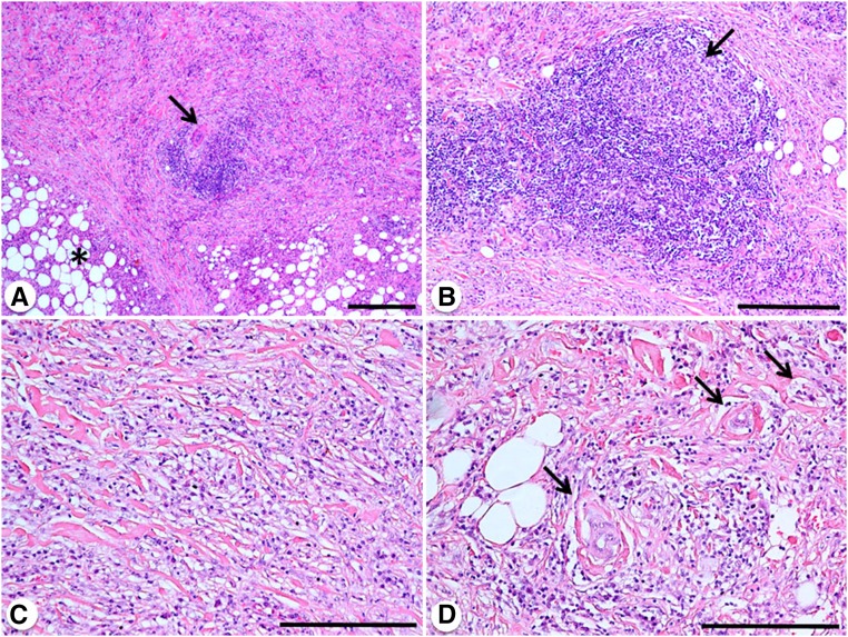
Figure 1.
Histopathology of idiopathic RPF. (A) Low-power magnification view of a retroperitoneal biopsy showing abundant and irregular fibrosis replacing normal retroperitoneal soft tissues (asterisk), and an inflammatory infiltrate organized in a lymphoid aggregate that is centered around a small retroperitoneal artery (arrow). (B) A lymphoid nodular aggregate with a clear (germinal) center (arrow) is visible. (C) Diffuse pattern of the inflammatory infiltrate, mainly consisting of lymphocytes and plasma cells that are diffusely interspersed within collagen bundles. (D) These collagen bundles form rinds around small retroperitoneal vessels (arrows). Hematoxylin and eosin (A–D). Original magnification, ×4 in A (bar is 0.5 mm); ×10 in B (bar is 300 μm); ×20 in C and D (bar is 300 μm).

