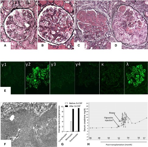
Figure 1.
Kidney biopsy findings in the native as well as the serial allograft biopsies. (A) Native kidney biopsy showing a glomerulus with expansion of the mesangial matrix and membranoproliferative features. (B) Second kidney allograft biopsy showing a glomerulus with largely mesangial proliferative features. (C) Third kidney allograft biopsy showing a glomerulus with expansion of the mesangial matrix and membranoproliferative features reminiscent of the pattern of injury observed in native kidney. (D) Fourth kidney allograft biopsy showing a glomerulus with circumferential cellular crescent (Jones methenamine silver). Original magnification, ×400. Notably, all allograft biopsies showed negative C4d staining along peritubular capillaries. Furthermore, circulating donor–specific antibodies, which were assessed only at the time of the third and fifth allograft biopsies using single–bead Luminex assay, were undetectable. (E) All native and allograft biopsies showed intense staining for γ2-heavy chain and λ-light chain with negative staining for γ1-, γ3-, γ4-heavy chains, and κ-light chain (fourth allograft biopsy; immunofluorescence). Original magnification, ×400. (F) Electron-dense deposits with 17-nm lattice–like parallel arrays (fourth allograft biopsy; electron microscopy). Original magnification, ×40,000. (G) Average number of intraglomerular lymphocytes, monocytes, and granulocytes assessed in the third and fourth allograft biopsies using immunohistochemical staining for CD3, CD68, and myeloperoxidase, respectively. (H) Change of serum creatinine in relation to filgrastim injection. G-CSF, granulocyte colony–stimulating factor.

