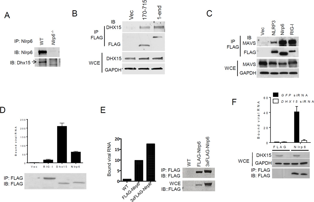
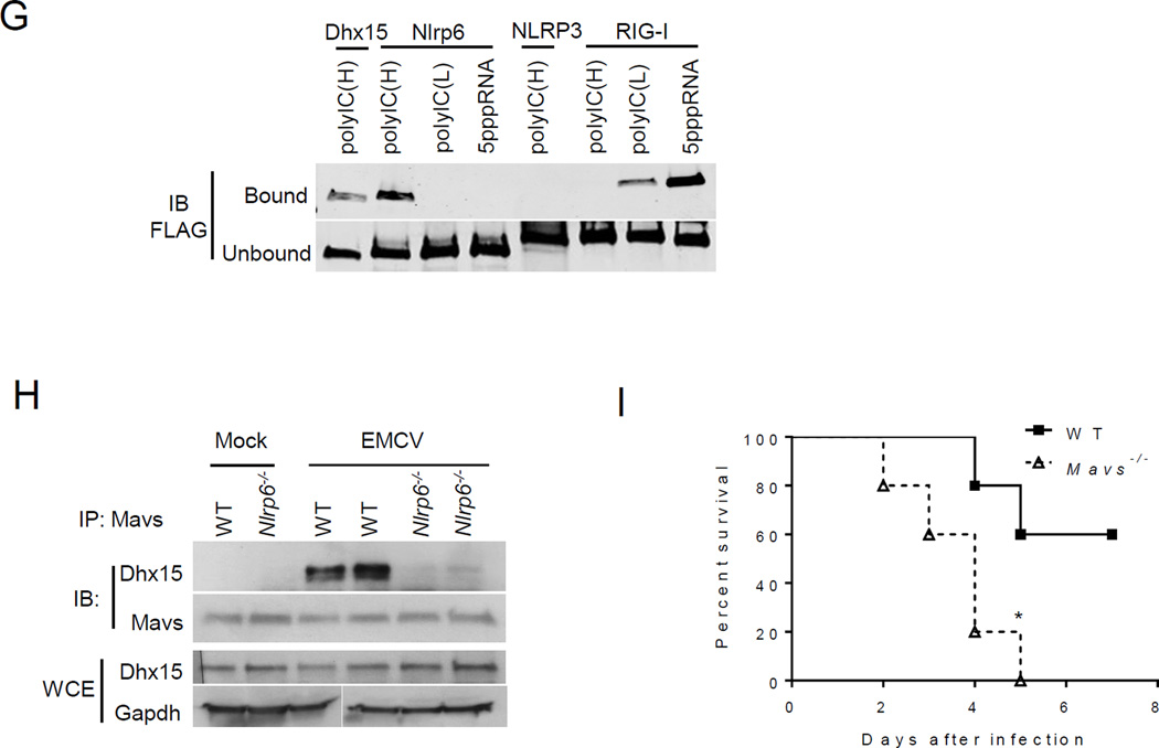
Fig. 2. Nlrp6 binds viral RNA via Dhx15.
(A) Co- immunoprecipitation (IP) of Nlrp6 with Dhx15 from WT and Nlrp6−/− mouse intestinal epithelial cells using an anti-Nlrp6 antibody. IB: immunoblotting. (B) Co-IP of FLAG-Nlrp6 NACHT+NAD (amino residues 170–715) and the full-length (1–end) with endogenous DHX15 from HEK293T cells overexpressing FLAG-tagged proteins using an anti-FLAG antibody. (C) Co-IP of FLAG-tagged proteins with endogenous MAVS from HEK293T cells as in (B). WCE, whole cell extract. RIG-I, retinoic acid inducible gene 1; GAPDH, glyceraldehyde 3-phosphate dehydrogenase. (D) Quantitative PCR analyses of viral RNA bound by FLAG-tagged proteins from EMCV-infected and FLAG fusion protein-expressing HEK293T cells. The data are presented as fold increase over vector (FLAG). (E) Binding of endogenous Nlrp6 to viral RNA. Left chart, quantitative PCR analyses of viral RNA bound by endogenous FLAG-Nlrp6 in IECs. Right panel: immunoblots of FLAG-Nlrp6 in WCE and IP. 3xFLAG-Nlrp6 denotes 3 FLAG motifs tagged to Nlrp6. (F) Quantitative PCR analyses of EMCV RNA bound by FLAG-Nlrp6 from GFP or DHX15 siRNA-treated HEK293T cells. The lower panel: immunoblots of WCE and IP. (G) Immunoblots showing FLAG-tagged proteins (purified from HEK293T) bound by biotin-labeled RNA analogues. PolyIC(H): high molecular weight (1.5–8kb), polyIC(L): low molecular weight (0.2–1kb). (H) Co-IP of Mavs with Dhx15 from IECs of WT and Nlrp6−/− mice infected with EMCV using a rabbit anti-Mavs antibody. (I) The survival curves of WT and Mavs−/− mice after oral infection with EMCV. N=5/group; *, p<0.05 (Log-rank test). The data are representative of at least two independent experiments.


