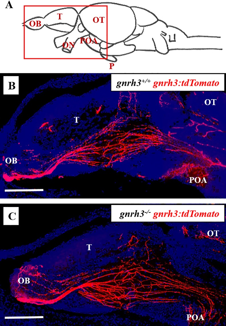
Fig 7. gnrh3-/- juveniles exhibit normal Gnrh3 neuronal migration.

In both gnrh3+/+ gnrh3:tdTomato (B) and gnrh3-/- gnrh3:tdTomato (C) fish, Gnrh3-tdTomato-expressing soma located in the olfactory region and ventral telencephalon project fibers that extend posteriorly toward the hypothalamus and pituitary stalk (pituitaries not shown; A). Z-stack images were taken at 20x magnification on a Leica Microsystems DMi8 confocal microscope with a resolution of 1024 x 1024 and a z-step size of 0.10. All images were analyzed and assembled with Image J and Adobe Photoshop. Scale bars = 100 μm. OB = olfactory bulb. T = telencephalon. OT = optic tectum. ON = optic nerves. POA = pre-optic area. P = pituitary.
