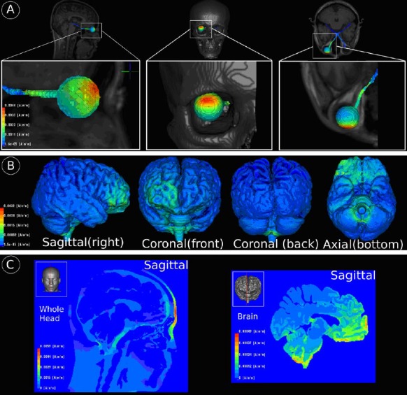
Fig 3.

Visualization of simulated electrical fields during rtACS: current density maxima on eye/optical nerve (A), brain tissue surface (B) and in the volume (C). Although four electrodes were used for treatment, they were used only one at a time. Therefore, the current flow simulation was done with only one electrode, representing all other electrodes. (A) Current density maxima of about 0.0044 A/m2 can be observed on the upper part of the outer eye surface that is closest to the stimulating electrode. Furthermore, the optical nerve of the stimulated eye also receives parts of the stimulating current density magnitude as currents enter the inner skull. (B) Local current density maxima can be found at frontal brain regions spatially located close to the stimulating anodal electrode. (C) Another area of locally increased current density can be found at the brain stem and lower cerebellum.
