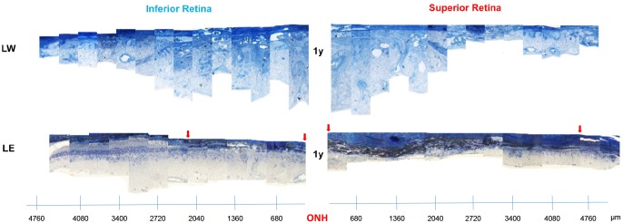
Fig 4. Representative reconstruction of the inferior (left) and superior (right) retina (composed of 12–13 consecutive histological segments of 75μm in width, each sectioned at every 340μm from the ONH to the ora serrate of each hemiretina) obtained from two different strains (LW and LE) of adult light-exposed rats over 1 year following light exposure.
Abbreviations: Optic nerve head (ONH), Lewis (LW) and Long Evans (LE). Red arrows indicate the portion of the retina devoid of photoreceptors.

