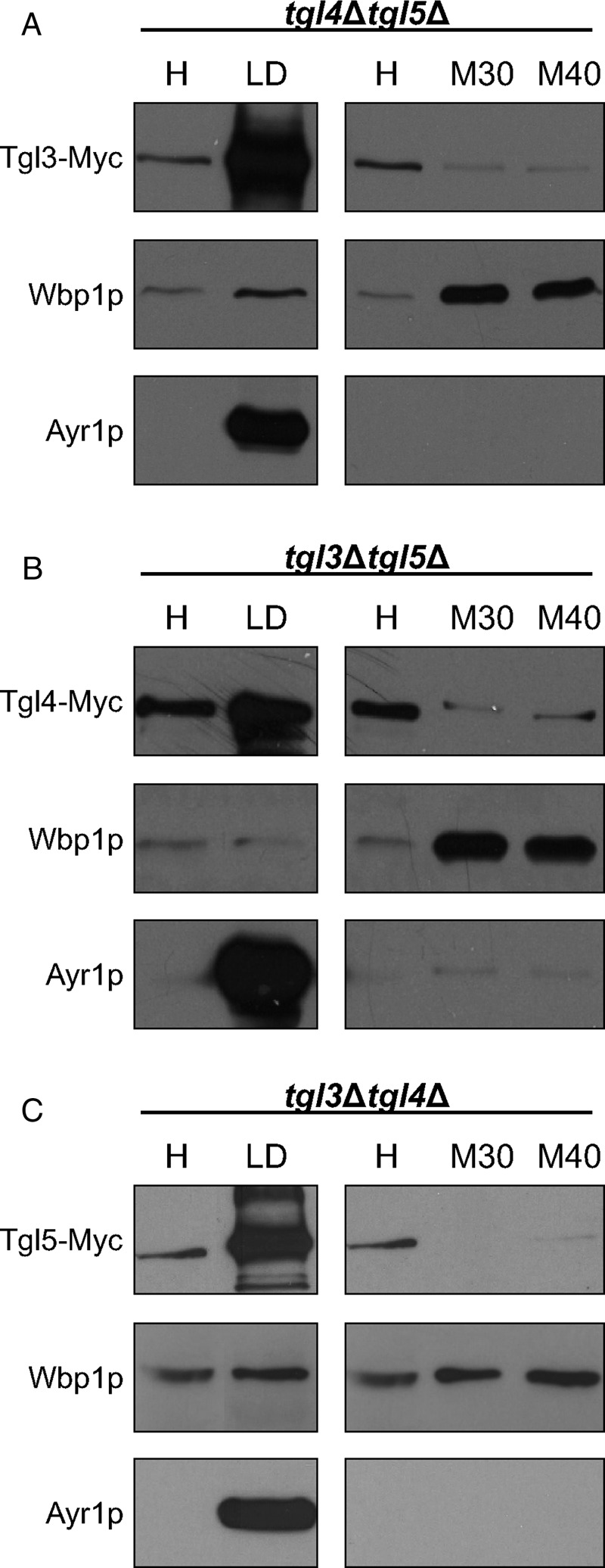
FIGURE 9:

Localization of Tgl3p, Tgl4p, and Tgl5p in the absence of counterpart lipases. Western blot analysis of (A) Tgl3-Myc in tgl4∆tgl5∆, (B) Tgl4-Myc in tgl3∆tgl5∆, and (C) Tgl5-Myc in tgl3∆tgl4∆ using samples of homogenate (H), 30,000 × g microsomes (M30), 40,000 × g microsomes (M40), and LD fractions is shown. Primary antibodies were directed against the Myc tag (Tgl3-Myc, Tgl4-Myc, and Tgl5-Myc), Wbp1p (ER marker), and Ayr1p (LD marker). Western blot analyses are representative of at least two independent experiments.
