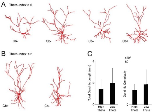
Author response image 2. Dendritic morphologies of L2 neurons.

(A) Reconstructed dendritic morphologies of the L2 neurons whichdisplayed theta-rhythmic spike discharges (theta- indices ≥ 5; see Figure 6E in the revised manuscript). (B) Reconstructed dendritic morphologies of the L2 neurons, whose spiking activitywas not rhythmically entrained by theta oscillations (theta-indices ≤ 5; see Figure 6E in the revised manuscript). (C) Total dendritic lengths (left bar graph) and dendritic complexity index (calculated as in Pillai et al., 2012; right bar graph) for theta-rhythmic (‘high-theta’) and non-theta -rhythmic neurons (‘low theta’) shown in A and B, respectively. Error bars represent SD. These differences were not statistically significant.
