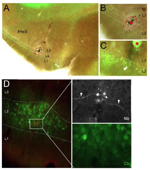
Author response image 3. Representative electrolytic lesions and ‘recording site’, which aided identification of PreS L2 in a subset of preliminary experiments.

(A) Parasagittal section trough PreS showing the reconstructed electrode track (dotted line) and a large electrolyticlesion (dotted circle) centered on PreS L2. Green, calbindin staining. (B) High-magnifications view of the electrolytic lesion shown in A. (C) High-magnification example of another electrolytic lesion (dottedcircle and asterisk) recovered at the expecteddistance from the recording site (end of the electrode track, indicate by the arrowhead). (D) Parasagittal section trough PreS stained for Neurobiotin (red) and calbindin (green), showing a representative recording site’ within L. Here, cell identification by juxtacellular labeling failed; however, cell debris and small portions of dendrites (rig t panels; arrowheads) could be observed at the labeling site within PreS L2.
