
FIGURE 2.
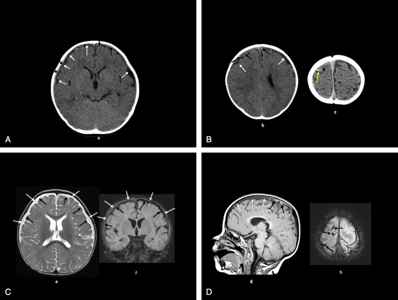
A, Patient 2: CT (a) shows bilateral mildly moderately large low-density inner subarachnoid collections (white arrows), small isohyperdense outer subdural collections (black arrows). B, Patient 2: CT (b, c) shows bilateral mildly moderately large low-density inner subarachnoid collections (white arrows, b), small isohyperdense outer subdural collections (black arrows, b), high-density cortical vein (yellow arrow, c). C, Patient 2: MRI axial T2 (e), coronal fluid attenuated inversion recovery (FLAIR) (f); bilateral moderately large T2 high-intensity, FLAIR low-intensity inner subarachnoid collections (black arrows) with T2 high-intensity, FLAIR isointense outer subdural collections (white arrows). D, Patient 2: MRI sagittal T1 (g), axial T2* (h: bilateral T1 isohypointense extracerebral collections (white arrows) with more recent T1 hyperintense, T2* hypointense hemorrhages, venous thromboses (black arrows).