
FIGURE 3.
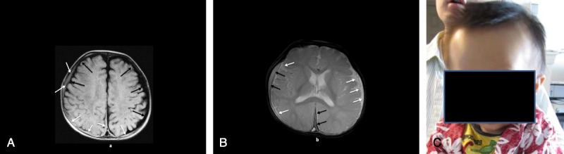
a, Patient 3: MRI axial T1 image (A) shows bilateral large lower-intensity subarachnoid spaces (black arrows) with superimposed higher-intensity bilateral convexity and interhemispheric subdural collections (white arrows). B, Patient 3: MRI axial T2* image (B) shows bilateral large high-intensity subarachnoid spaces (white arrows) with superimposed lower-intensity right convexity and interhemispheric subdural collections (black arrows). C, Patient 3: at 9 months of age showing frontal bossing and macrocephaly.