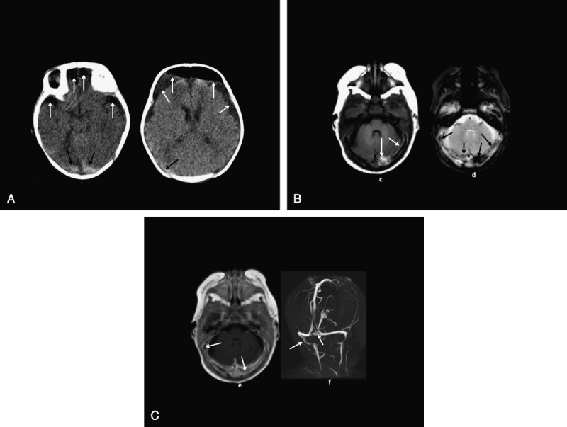
FIGURE 5.

A, Patient 6: CT (A, B) shows postsurgery frontal extracerebral low-density air-fluid collections (white arrows) and high-density hemorrhages or thromboses along the tentorium (black arrows). B, Patient 6: MRI axial T1 (C) and T2* (D) images show T1 high plus T2* low-intensity hemorrhages and/or thromboses along the transverse dural venous sinuses (arrows). C, Patient 6: MRI axial gadolinium T1 (E) and posterior MRV (F) images show asymmetric left transverse and right sigmoid dural venous sinus flow gaps (arrows) consistent with thromboses.
