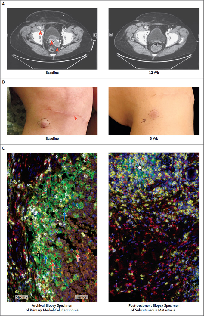
Figure 3 (facing page). Response to Pembrolizumab in a Patient with Stage IV Merkel-Cell Carcinoma.
This 69-year-old woman received a diagnosis of a primary cutaneous lesion on the right knee and was treated with wide local excision, sentinel lymph-node biopsy, and inguinal lymph-node dissection in November 2013. Recurrent Merkel-cell carcinoma developed in September 2014, with a pelvic mass measuring 11 cm by 7 cm by 14 cm, which was associated with worsening lymphedema and moderate-to-severe right hydroureterone-phrosis requiring a ureteral stent. The patient received radiation therapy to the pelvic mass but in January 2015 was found to have new peritoneal and lymph-node metastases (Panel A, red arrows), as well as several subcutaneous metastases on the right thigh and just below the site of excision of the primary tumor (Panel B; red arrow indicates the site of previous excision of the primary tumor, just below the knee). As shown, these metastatic sites regressed rapidly during anti–programmed death 1 (PD-1) therapy. Also shown are the results of pathological analysis of the primary tumor (Panel C, left) and adjacent post-treatment subcutaneous metastasis (Panel C, right) with multispectral immunohistochemical analysis. Orange indicates Merkel carcinoma cells expressing neuron-specific enolase, yellow CD8+ T cells, red CD68+ macrophages, white PD-1, green the PD-1 ligand PD-L1, and blue nuclear DNA stained with 4′,6-diamidino-2-phenylindole (DAPI). Analysis of the archival biopsy specimen shows an immune infiltrate that is most intense at the tumor–stromal interface, including CD68+ macrophages and CD8+ T cells infiltrating the tumor parenchyma. PD-1 is expressed on 56% of CD8 cells in this microscopic field. PD-L1 is expressed on tumor cells (10% of tumor cells in this field, blue arrows) and macrophages (43% of macrophages in this field, red arrows) and is seen immediately adjacent to PD-1+ lymphocytes. Analysis of the post-treatment biopsy specimen shows a diffuse immune-phagocytic infiltrate and no evidence of residual tumor. The immune infiltrate includes CD68+ macrophages and CD8+ T cells, with an early lymphoid aggregate (white star) where PD-1 and PD-L1 expression is observed.

