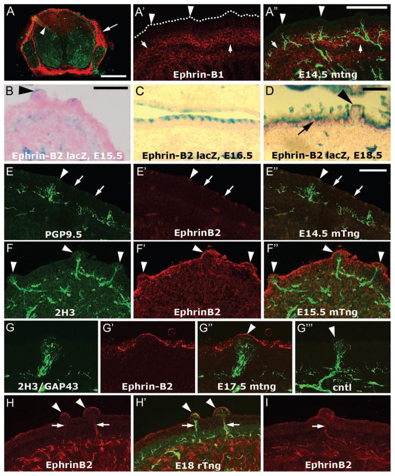
Figure 1.
Localization of ephrin-B1 and ephrin-B2 in and near the dorsal lingual epithelium during target penetration by sensory afferents. A–A″. TSA Plus-enhanced monoclonal ephrin-B1 immunostaining. A. We labeled sections of mouse E12.5 spinal cord as a positive control for ephrin-B1 immunostaining. Anti-ephrin-B1 (red) strongly labels the mesenchyme surrounding the spinal cord and DRG (arrow), and moderately labels dorsal radial glia (arrowhead). PGP9.5 staining is shown in green. A′–A″. Ephrin-B1 staining (red) was found in the tissue subjacent to the epithelium (A′, arrows) and is difficult to detect in the epithelium, including the fungiform papillae (arrowheads). The oral surface of the epithelium is marked by dots. PGP9.5 was used to label axons (green). Note that the brightness and contrast of the ephrin-B1 staining has been enhanced, it is dimmer than the positive control staining of the dorsal radial glia in the spinal cord (Fig. 1A, arrowhead). B–D. LacZ staining in ephrin-B2lacZ mouse embryos. A low level of ephrin-B2 lacZ staining was often detected at the serosal surface of fungiform papilla epithelium at E15.5 (B, arrowhead) and E16.5. In addition, more intense periodic lacZ staining was found in the posterior half (E15.5) and 3/4ths (E16.5, C) of the apical aspect of the dorsal lingual epithelium. By E18.5 (D), the ephrin-B2 lacZ staining was continuous along the dorsal epithelium of the tongue (arrow), but more apically restricted within fungiform papillae (arrowhead). E–E″. Intermittent anti-ephrin-B2 labeling (red, arrows) was evident along the oral surface of the lingual epithelium, including fungiform papilla epithelium (arrowheads) as early as E14.5 in mouse. F–F″. In E15.5 mouse tongue (F″), anti-ephrin-B2 staining (F′) was typically continuous along the dorsal lingual epithelium, including fungiform papilla epithelium (arrowheads, F–F″), and did not colocalize with 2H3 anti-neurofilament nerve staining (F). G–G‴. Anti-ephrin-B2 labels the serosal surface of a murine E17.5 fungiform papilla (G′, arrowhead in G″) that is substantially innervated (G). A combination of two monoclonal antibodies, 2H3 and anti-GAP43 was used to detect axons. In the negative control in which no primary antibody was applied (G‴), no staining was observed in the red channel in fungiform papilla (arrowhead). H–I. In E18 rat tongue, however, ephrin-B2 labeling (red) was present in fungiform papilla epithelium (arrowheads, H,H′) but also colocalized with 2H3/GAP43 labeled afferents (H′, green). This staining was also observed when anti-ephrin-B2 was the only antibody used to label the tongue section (I), showing that it is not due to “bleed through” or adventitious excitation of the alexafluor 488 that we typically use to detect 2H3/GAP43 in the axons. Calibration bar in A = 250 μm; bar in A″ = 100 μm; bars in B, D, and E″ = 50 μm. Bar in B applies to C and bar in E″ applies to E–I.

