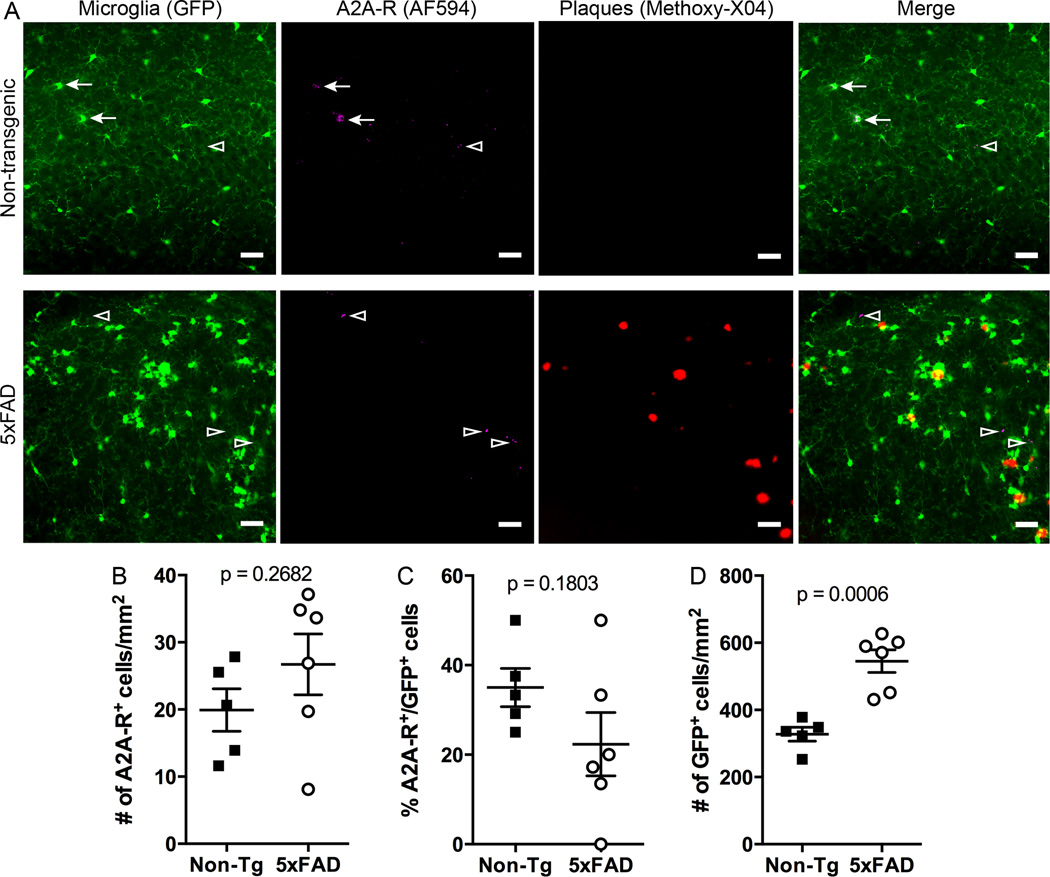
Figure 4. Adenosine A2A receptor expression in 5xFAD mice.
5xFAD:mg-GFP and non-Tg mice were injected with Methoxy-X04 and brains were isolated and fixed 24 hr later. A. Brain sections (40 µm) were stained for A2A receptor and imaged on a confocal microscope to obtain GFP (microglia, shown in green), AlexaFluor-594 (A2A receptors, shown in magenta) and Methoxy-X04 (plaques, shown in red) signals. Merged images show A2A receptor-positive microglia in the hippocampus of both non-Tg and 5xFAD mice. Scale bar: 30 µm. Arrows point to GFP-positive cells that appear to colocalize with A2A receptors. Arrowheads point to A2A receptor-positive cells that do not colocalize with GFP-positive microglia. B-D. Quantification of A2A receptor-positive cells. The total number of A2A receptor-positive cells in the hippocampus is not significantly different between 5xFAD and non-Tg mice (B). The number of A2A receptor-expressing microglia is not significantly different between non-Tg and 5xFAD mice (C), despite the overall higher number of microglia in 5xFAD mice (D). Statistics: Student’s t test.

