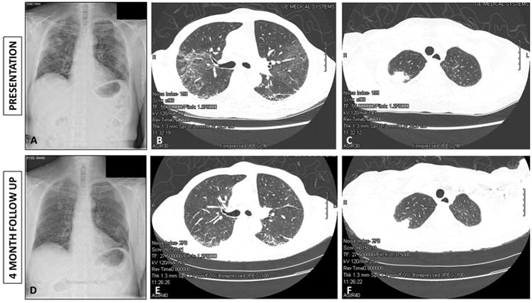
Figure 2.

A: Chest radiograph at time of presentation with hemoptysis showing apical lesions with central cavitation. B: Thoracic computed tomography at time of presentation demonstrating fibrotic changes with honey combing and traction bronchiectasis. C: Thoracic computed tomography demonstrating right apical lung lesion with central cavitation. D: Follow-up Chest radiograph four months after commencing induction therapy with steroids and rituximab demonstrating interval improvement in the lung lesions. E: Follow-up thoracic computed tomography demonstrating interval improvement in the interstitial lung disease. F: Follow-up thoracic computed tomography demonstrating partial resolution of the apical cavitating lesions four months after institution of induction therapy with steroids and rituximab.
