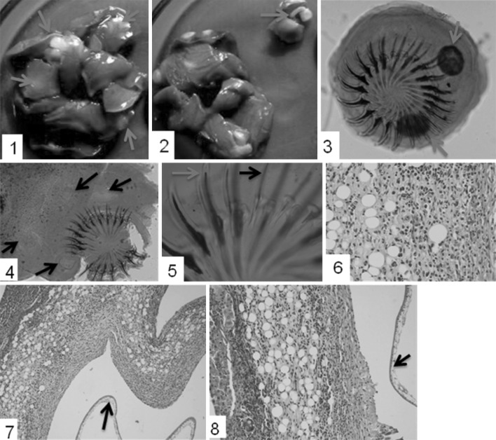
Fig. 1.
1 Multifocal hepatic cysts (arrows) involving all the lobes. 2 Incised section of the cyst depicting strobilocercus/metacestode (arrow). 3, 4 Mature scolex of metacestode with two distinct rows of large and small hooks with presence of lateral suckers (arrows). 5 Comparative representation of large (blue arrow) and small (black arrow) hooks. 6 Inflammatory reaction around the cyst with eosinophils and fatty changes. H E × 40X. 7, 8 Hepatic lesions possessing fibrous tissue, inflammatory cells and fatty change or metaplasia around the parasitic cyst. There is presence of healthy tissue, zone of inflammation and cyst wall (arrow). H E × 10X. (Color figure online)

