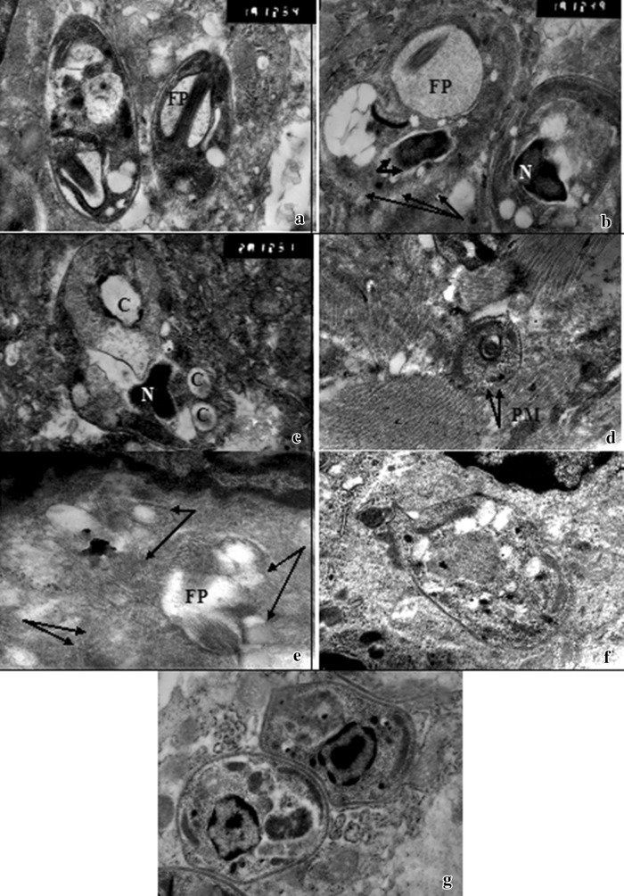
Fig. 2.
Transmission electron micrograph (TEM) of cutaneous lesions of infected treated mice a, b, c Infected mice treated with miltefosine (group II) showing a L. amastigotes with flagellar knot inside dilated flagellar pocket (FP) (15,000×). b L. amastigotes with extensive dilatation of flagellar pocket (FP), disintegration of plasma membrane (arrows), abnormal nuclear chromatin condensation (N), and absence of nuclear membrane (thick short arrows) (15,000). c L. amastigotes with homogenous nucleus (N), cytoplasmic vaculization, and multiple membrane bound cavities (C) (20,000×). d, e, f Infected mouse treated with azithromycin (group IIIa) showing d L. amastigotes with disintegration of plasma membrane (PM) (arrows) (7,500×). e L. amastigotes with ill defined parasite contour and content(arrows) but with evident flagellar pocket (FP) (15,000×). f L. amastigotes with cytoplasmic vacuolization (13,000×). g L. amastigotes of infected untreated control group

