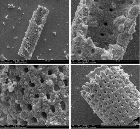
Fig. 1.

(a, up left) Single micrometric diatom frustule. (b, up right) Au nanoparticle covers homogeneously the outer and inner surfaces of the frustule. (c, down left) An enlargement of outer surface with pore size measurement. (d, down right) A smaller micrometric frustule. All images are relative to 48 h of Au deposition time
