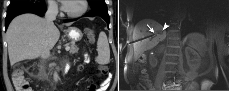
Fig. 1.
A 63-year old patient with a small lesion in liver segment VII was referred for MRI-guided biopsy due to lesion invisibility on CT (left). With MRI (VIBE) the lesion (arrowhead) was clearly depicted without contrast agent administration (right). The double oblique access path was 131 mm. With the help of the navigation device a coaxial needle is inserted before the lesion. Through the needle a 16 G semiautomatic biopsy gun is placed. The Tru-Cut® tip of the biopsy gun is recognizable (arrow) at the border of the lesion. The histology analysis revealed a non-Hodgkin’s lymphoma

