Fig. 4.
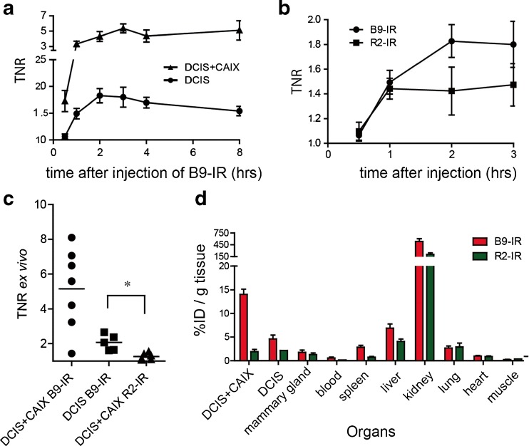
Optimal imaging with B9-IR nanobody 2 h post injection. a Mean TNR of CAIX-overexpressing tumors (DCIS + CAIX, n = 10), and DCIS tumors (n = 10) determined during the first 8-h post injection of B9-IR nanobody. Error bars represent SEM. b Mice xenografted with DCIS tumors were injected with 50 μl B9-IR (10 mice) or R2-IR (4 mice) non-relevant control nanobody, mean TNR values were determined at indicated time points. c DCIS + CAIX (7 mice) and DCIS (7 mice) tumors after injection with 50 μg B9-IR, and DCIS + CAIX tumors (n = 6) after injection with R2-IR non-relevant control nanobody. Single values of intra-operative TNRs were determined 3 h post injection. Bar represents the mean (*p = 0.04). d For a biodistribution assay, mice (n = 9) were injected with B9-IR or R2-IR non-relevant control nanobody. Tumors and organs were collected 3 h post injection. Error bars represent SEMs.

