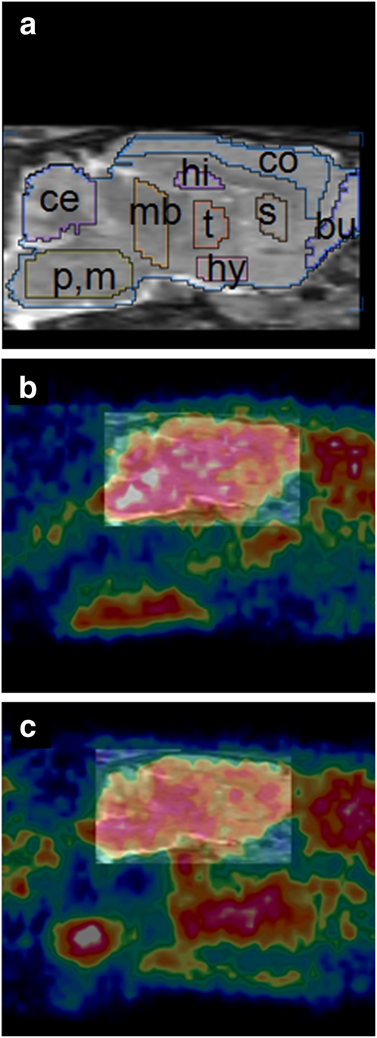
Fig. 1.
Images of rat brain (sagittal views, animals anesthetized with isoflurane). a MRI template. Brain regions are indicated by the following abbreviations: bu bulbus olfactorius, ce cerebellum, co cortex, hi hippocampus, hy hypothalamus, mb midbrain, p,m pons and medulla, s striatum, and t thalamus. b MicroPET image of young rat (age 3 months) superimposed on the MRI template. c MicroPET image of old rat (age 18 months) superimposed on this template. Frames from 22 to 74 min were summed (since early frames may contain a significant contribution of radioactivity in the blood pool), and a plane was chosen about 1.6 mm from the midline. Note the high uptake in the pons and medulla of the young rat whereas the distribution of radioactivity in the brain of the old rat is more uniform.

