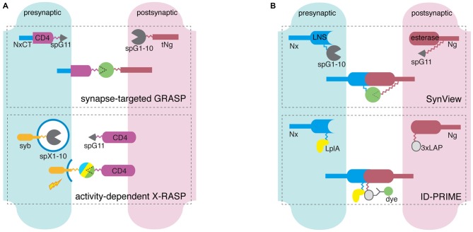
Figure 2.
Scheme of dual component synapse detectors. (A) GFP reconstitution-dependent molecular tools detect pre- and postsynaptic membrane apposition through the reconstitution of spGFP fragments each attached to the extracellular ends of membrane protein carriers. Synapse-targeted GFP Reconstitution Across Synaptic Partners (GRASP) utilizes a presynaptic component of spGFP11 (spG11, circular wedge) linked to a CD4-neurexin-1β C-terminus (NxCT) fusion through flexible peptide linkers (gray zigzag), and a postsynaptic component of spGFP1-10 (spG1-10) and truncated neuroligin-1 (tNg) connected with a linker. SpG11 and spG1-10 unite at synapses, not random membrane contacts. In activity-dependent X-RASP the presynaptic split-XFP1-10 (spX1-10) is fused to SNARE proteins such as synaptobrevin (syb), and unites with CD4-bound spG11 when presynaptic activity triggers vesicle release. (B) Neurexin (Nx)-neuroligin (Ng) interaction-dependent molecular tools detect individual synapses through fluorescent protein (FP)-based (SynView) and enzymatic reaction-based (Interaction-Dependent Probe Incorporation Mediated by Enzymes, ID-PRIME) direct visualization of Nx-Ng interaction. In SynView, spG11 is inserted between the esterase and proximal extracellular domain of Ng, and spG1-10 in the middle of the LNS domain of Nx. GFP reconstitutes when Nx and Ng unites. ID-PRIME uses the identical set of synaptic protein carriers, but tandem LAP tags (3xLAP) and lipoic acid ligase A (LplA) are attached to C-terminus of Ng esterase domain and Nx LNS domain, respectively. Nx-Ng interaction initiates LplA-mediated lipoic acid tagging to 3xLAP, and signals useful for imaging originate from antibody-fluorophore conjugates.

