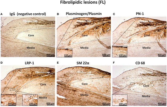
Figure 3.
Cellular localization of plasminogen/plasmin, PN-1, LRP-1, SM 22α, and CD68 in fibrolipidic lesions (FL). Serial sections of FL. IgG (negative control, A). Plasminogen/plasmin staining (B). Immunosignal of PN-1 (C). Immunostaining of LRP-1 (D). Smooth muscle cell marker staining (SM 22α, E). Phagocytosis marker (CD 68) staining (F).

