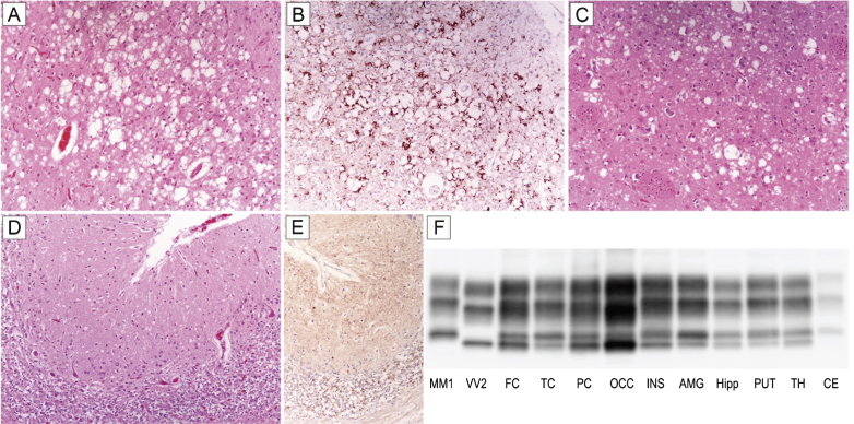
Fig.1.
Distinctive histopathology and regional distribution of PrPSc in patient #16 (MM 2C+1). (A) Spongiform change characterized by large, confluent vacuoles (H&E stain) and (B) perivacuolar and coarse PrP deposits (PrP immunohistochemistry) in the occipital cortex; (C) mixed type of spongiform change with large, confluent vacuoles intermixed with smaller vacuoles in the striatum (H&E stain); (D) spongiform change characterized by small, fine, microvacuoles (H&E stain) and (E) synaptic pattern of PrP deposition in the cerebellum (PrP immunohistochemistry). All pictures (A-E) have the same magnification (×100); F) PrPSc typing by western blot showing the co-occurrence of protein types 1 and 2 in all regions but the cerebellum, where only a relatively low amount of type 1 is seen. While the amount of PrPSc type 2 is higher than that of type 1 in most samples from the cerebral cortex, subcortical gray matter structures show either a similar amount of the two proteins or a predominant accumulation of type 1.

