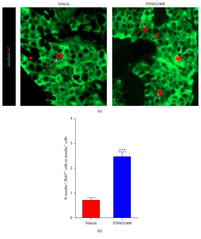
Figure 5.
(a) Immunofluorescent staining for insulin (green color) and Ki67 (red color) in human islet grafts from PSN632408- and vehicle-treated mice with normoglycemia at 4 weeks after transplantation. White arrows show insulin/Ki67 copositive cells in human islet grafts. (b) Percentage of insulin/Ki67 copositive cells among total insulin positive cells in human islet grafts. The average percent of insulin/Ki67 copositive cells among total insulin positive cells was significantly higher in PSN632408-treated mice (n = 9) compared with vehicle-treated mice (n = 4) (∗∗∗ P < 0.001).

