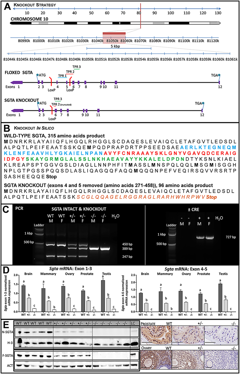
Figure 1. Generation of Mice Null for Small Glutamine-rich Tetratricopeptide Repeat-containing Protein Alpha (SGTA).
(A) Genomic structure of the mouse Sgta gene floxed at exons 4 and 5, and schematic of the resultant knockout product. (B) Protein sequence of wild-type SGTA, with TPR1, 2 and 3 highlighted in blue, red and green, respectively, and effect of exons 4–5 (amino acids 271–458) deletion leading to a 96 amino acid product. (C) PCR analyses confirming Sgta deletion in heterozygous (+/−) and homozygous (−/−) Sgta-null mice using ‘SGTA knockout’ primers, and the presence of Sgta in wild-type (WT) and Sgta heterozygous (+/−) mice, using ‘SGTA intact’ primers and finally the removal of Cre Recombinase (Cre) in a subset of Sgta+/− mice (L, ladder; bp, base pairs; H2O, water control; M, male; F, female; N.B. ladder’s two brightest bands at 1000 (upper) and 500 (lower) bp). (D) Quantitative Real Time PCR confirming homozygous Sgta mutation ablated Sgta mRNA compared to Sgta+/− and WT, while heterozygous Sgta mutation significantly diminished Sgta mRNA compared to WT. Analyses were conducted in brain, mammary, ovary, prostate, and testis tissues with primers spanning in either Sgta exon 1–3 and Sgta exon 4–5. Means (n = 8/group) with different letters are significantly different, P < 0.001. (E) Western blots (brain) and immunohistochemistry (prostate, ovary) confirming loss of SGTA protein in homozygous Sgta-null (−/−) tissue and lowered SGTA protein expression in heterozygous Sgta-null (+/−) tissue (LC, loading control; N-SGTA, probed with ‘N-terminal’ SGTA antibody; F-SGTA, probed with ‘Full length’ SGTA antibody; H-3, Histone H-3; ACT, Actin). For Western blots, representative cropped blots are depicted, with all blots run under the same experimental conditions. Scale bars represent 50μm.

