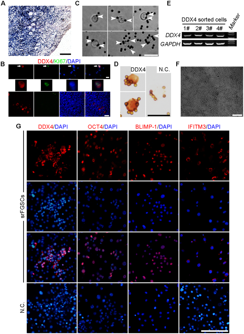
Figure 1. Isolation and characterization of human FGSCs from reproductive-age ovarian cortex of patients.
(A) Representative histological appearance of adult human ovarian tissue used for the isolation of srFGSCs. (B) Representative images of DDX4 (red) and KI67 (green) in putative FGSCs (upper panel) and oocytes (lower panel) in human ovarian cortex. The image of putative FGSC (white arrows) is enlarged in middle panel. (C) Representative images of DDX4+ cells from reproductive-age ovarian cortex. White arrowheads indicate the magnetic beads. (D) Immunocytochemical analysis of DDX4 expression in srFGSCs using the antibody against the C-terminus of protein. (E) RT-PCR analysis of DDX4 sorted cells. Lane 5, 100-bp DNA markers. GAPDH is sample loading control. (F) A representative morphology of srFGSCs at passage 31. (G) SrFGSC line was detected by immunofluorescence analysis with the antibodies against DDX4, OCT4, IFITM3, and BLIMP-1. Negative control (N.C.) in (D,G) are the omission of the primary antibody. Full-length gels in E are presented in Supplementary Fig. 5. Scale bars: 10 μm (upper panel of B), 20 μm (C,D), 50 μm (A, lower panel of B,G), 100 μm (F).

