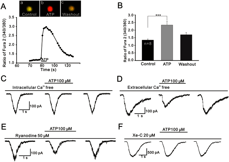
Figure 6. Calcium relevance of ATP-induced suppression of glycine currents.
(A) A continuous recording of [Ca2+]i in a GC, represented by the ratio of fura-2 AM fluorescence at 340 nm and 380 nm (340/380). Application of ATP dramatically increased [Ca2+]i in a reversible manner. Three CCD images of an another GC loaded with fura-2 AM were taken before (a) and 10 s following ATP perfusion (b), and after washout (c). (B) Bar chart showing the ATP-caused changes in [Ca2+]i in GCs. ***P < 0.001 vs. control. (C) Representative recordings from an OFF-GC, showing that during internal infusion of Ca2+-free solution (containing 10 mM BAPTA), ATP failed to suppress the glycine current. (D) Representative recordings from an OFF-GC, showing that ATP still suppressed the glycine current in Ca2+-free extracellular solution (containing 1 mM EGTA). (E,F) Current traces of two OFF-GCs, showing that during the internal infusion of ryanodine (50 μM) (E), but not Xe-C (20 μM) (F), the ATP suppression effect on glycine current was seen.

