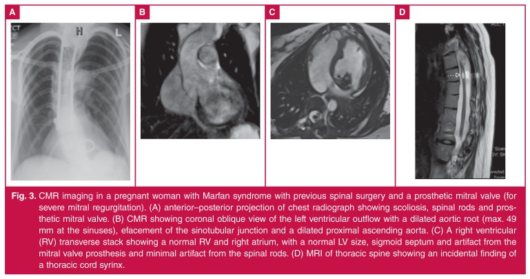
Fig. 3.
CMR imaging in a pregnant woman with Marfan syndrome with previous spinal surgery and a prosthetic mitral valve (for severe mitral regurgitation). (A) anterior–posterior projection of chest radiograph showing scoliosis, spinal rods and prosthetic mitral valve. (B) CMR showing coronal oblique view of the left ventricular outflow with a dilated aortic root (max. 49 mm at the sinuses), efacement of the sinotubular junction and a dilated proximal ascending aorta. (C) A right ventricular (RV) transverse stack showing a normal RV and right atrium, with a normal LV size, sigmoid septum and artifact from the mitral valve prosthesis and minimal artifact from the spinal rods. (D) MRI of thoracic spine showing an incidental finding of a thoracic cord syrinx.

