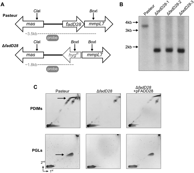
Figure 1. Construction of a PDIM/PGL deficient strain of BCG-Pasteur.
(A) Genomic organization of WT (Pasteur) and ∆fadD28 strains. Dashed lines indicate products of restriction digestion with ClaI and BoxI. (B) Southern blot analysis. Chromosomal DNAs isolated from WT and three randomly picked ∆fadD28 clones were digested with ClaI and BoxI and blotted with a 500 bp probe of fadD28, which yielded a 3.5 kb and 1.8 kb fragment, respectively, and agreed with prediction (A). (C) 2D-TLC analysis of PDIMs and PGLs. For PDIM analysis, apolar lipids were developed with petroleum ether/ethyl acetate (98:2 v/v, 3 times) in the first dimension (1st) and petroleum ether/acetone (98:2, v/v) in the second dimension (2nd). Lipids were visualized by charring with 5% phosphomolybdic acid. For PGL analysis, the apolar lipid extract was developed with chloroform/methanol (96:4, v/v) and toluene-acetone (80:20, v/v), followed by charring with α-naphthol. PDIMs, phthiocerol dimycocerosates; PGLs, phenolic glycolipids.

