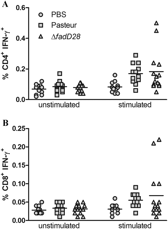
Figure 3. The loss of PDIMs/PGLs does not affect production of IFN-γ.

Intracellular cytokine staining analysis of IFN-γ production by (A) CD4+ and (B) CD8+ T-cells. C57BL/6 mice were immunized subcutaneously with the WT BCG-Pasteur, ∆fadD28, or PBS/0.01% Tween 80. At 9 weeks post-vaccination, mice were sacrificed and splenocytes were harvested. Splenocytes were incubated with or without PPD for 24 hr followed by staining for T-cell surface markers (CD3-PE, CD4-FITC, CD8a-PercyPCy5.5) and intracellular IFN-γ (IFN-γ-APC). Samples were analyzed by BD FACSCaliburTM and FlowJo© Software. Pooled results from two independent experiments; each data point represents one mouse.
