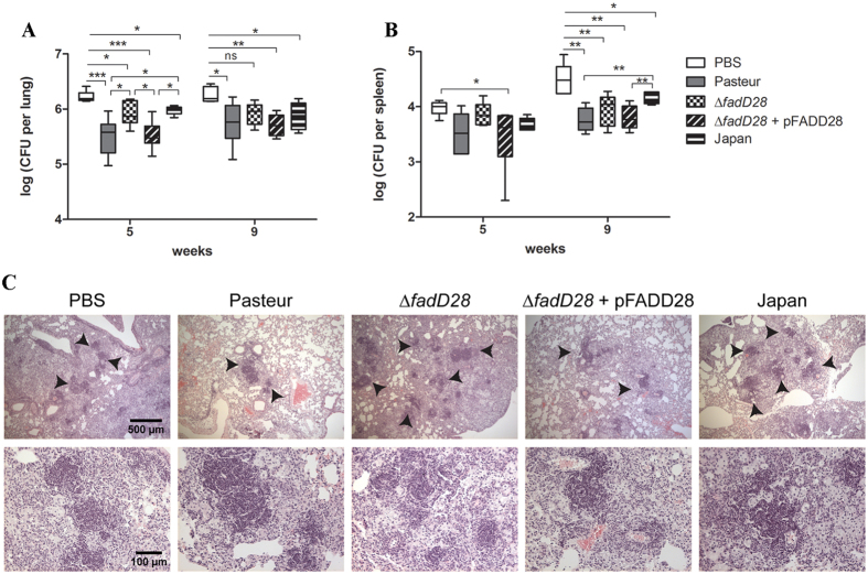
Figure 4. Loss of PDIMs/PGLs reduces BCG-mediated protection against M. tb in mice.
BALB/c mice were vaccinated subcutaneously with ~105 CFU of the BCG strains or PBS as a control. At 8-weeks post vaccination, mice were aerogenically challenged with M. tb. Mice were sacrificed at 5 and 9 weeks post-challenge and organs were examined for bacterial burden and pathology. The M. tb burden in the (A) lungs and (B) spleen was shown (6 mice per group per time point; data were plotted as box-whiskers in which the whiskers represent the minimum and maximum of all data. *p < 0.05; **p < 0.01; ***p < 0.001; one-way ANOVA, Tukey’s post hoc test). (C) Histological analysis of lung sections from mice in each group at 9 weeks post-challenge. Samples are stained with H&E. Arrows indicate regions of granuloma-like lesions. Top row is 50x magnification; bottom row is 200x magnification.

