
Fig. 8.
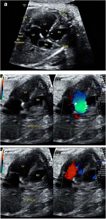
Sonographic images of a 24-gestational-week fetus in which VSD was falsely suspected. In the 4CV, a small echo drop-out was visualized at the perimembraneous septum, as indicated by the white arrows (a). This was at times easily considered to be a VSD, whereas it was actually an artifact. Color Doppler helped to resolve this problem. No flow signals were identified across the septum in either systolic (b) or diastolic (c) phases