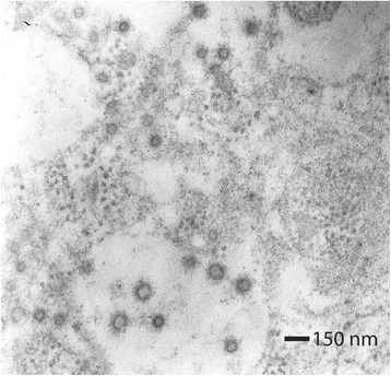
Fig. 3.

Electronic transmission photography of an enterocyte’s cytoplasm. Numerous viral particles measuring 75–83 nm in diameter are observed. These viral particles possess a membrane with numerous slightly electrodense projections that are 20 nm in length (arrow). Adjacent to these viral particles, clusters of ribosomes (inset) are appreciated. Contrast technique with uranyl acetate and lead citrate. 50,000× magnification
