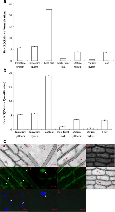
Fig. 2.

qRT-PCR analysis of the expression of PdRanBP in different vascular tissues and organs of P. deltoides, and detection of immediate and stable expression of GFP-tagged PdRanBP. a, b qRT-PCR analysis of PdRanBP expression in the vascular tissues and other organs of P. deltoides during secondary cell wall development. Aliquots of 1000 ng total RNA were reverse-transcribed into cDNA. The signals were normalized to the constitutively expressed poplar α-tubulin (TUA1) (a) and Ubiquitin (UBQ1) (b) genes. The values are the mean ± standard error (SE) of three replicates. PdRanBP was predominantly expressed in the leaf buds, immature xylem and immature phloem of P. deltoides. c Nuclear localization of EGFP-PdRanBP fusion protein in onion epidermal cells. Dark-field images were captured for green fluorescence (e and f), GFP-only control (g) and the corresponding bright-field images for e, f, g are a, b, c. Bright-field images (h) were captured for cell morphology, and the corresponding dark-field images for h is d. i and j, Nuclei counterstained with 4′, 6-diamidino-2-phenylindole (DAPI); the corresponding GFP-only control images of i and j are shown in k. The scale bars are 200 μm in a, c, d, e, g, h, i and k, and 800 μm in b, f, and j
