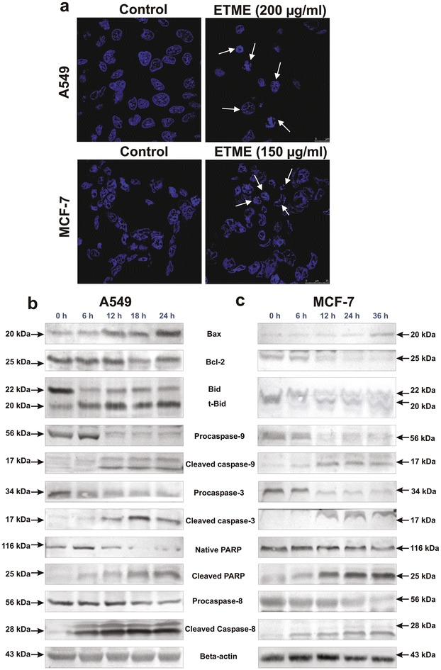
Fig. 3.

Observation of DNA fragmentation and westerblot analysis of apoptotic proteins in ETME treated A549 and MCF-7 cells. a The fragmented DNA of nuclei were bind with DAPI and observed under a confocal microscope (630×). The white arrows indicate cells with fragmented DNA of nucleus after the treatment of ETME. b Effect of ETME on apoptotic proteins in A549 (200 µg/ml) cells and c MCF-7 (150 µg/ml) cells analysed by immunoblotting after various time intervals. Expression of β-actin was set as a protein loading control
