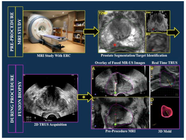
Figure 1.
Workflow of magnetic resonance imaging (MRI)/ultrasound (US) fusion-guided prostate biopsy. Red asterisks indicate a right medical apical lesion as seen on T2W=T2 weighted imaging (green contour shows MRI prostate segementation), DWI=diffusion weighted image and DCE=dynamic contrast enhanced image sequences. After 2D TRUS sweep of the prostate, the acquired images are used to create a 3D US reconstruction. This 3D rendering is co-registered (elastic/rigid registration) with the MRI dateset. MR-US co-registered images are co-displayed above (A) and beside (A,B) each other. 3D rendering of the prostate (with overlaid needle core location) generated after sampling and tracking. TRUS= transrectal ultrasound; 2D= 2-dimensional; 3D= 3-dimensional.

