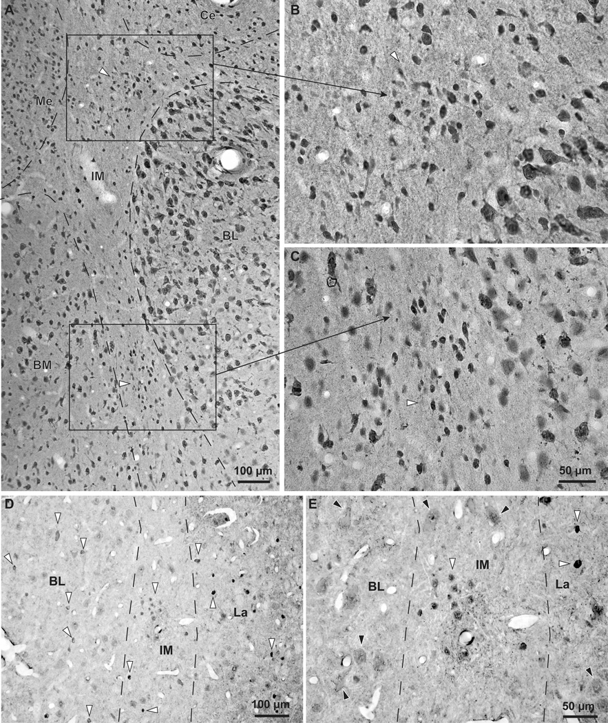
Figure 4.
The IM clusters in the rhesus monkey brain. NeuN and GABA immunohistochemistry in the IM of the rhesus monkey amygdala. A–C, Coronal brain section stained for NeuN through the amygdala. A, Low magnification shows the demarcated IM, including some clusters (white and black, silhouette arrowheads). B–C, IM clusters are seen at higher magnification. Note the small size of IM neurons compared to neurons in BL and BM. D–E, Isolated inhibitory neurons and small IM neuron clusters (white and black, silhouette arrowheads) are labeled with GABA. BL and La also contain inhibitory GABAergic neurons that can be darkly or lightly stained (white and black, silhouette arrowheads). In contrast, pyramidal neurons in BL and La are larger in size, often have pyramidal shape, and have very light, background levels of staining in their cytoplasm (black arrowheads in E). In most cases pyramidal neuron somata appear to be surrounded by darkly-stained GABAergic axon terminals that form perineuronal complex basket formations, previously described on pyramidal neurons in cortical and amygdalar areas of many species, including monkeys and humans (black arrowheads in E). Scale bar in C applies to B and C.

