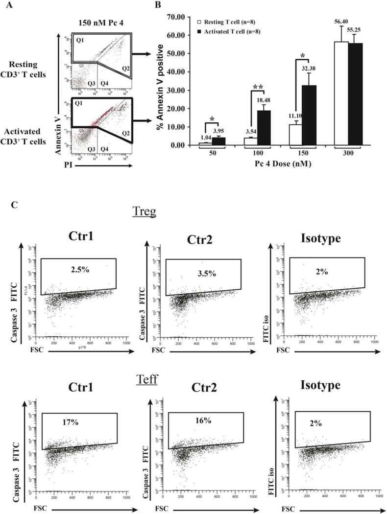
Fig 2. Activated T cells exhibit greater susceptibility to Pc 4-PDT induced cell death.

Activated or resting T cells were incubated with varying doses of Pc 4 (0–300 nM) in complete medium for 2 h, followed by irradiation with 200 mJ/cm2 of red light (λmax = 675 nm). After 4 h, cells were harvested and stained with fluorescein isothiocyanate (FITC)-labeled anti-Annexin V mAb and PI. Flow cytometric analysis was performed using the BD FACSAria flow cytometer. Baseline levels of cell death (~7% and ~15% in Q1 and Q2, respectively) were subtracted for each sample. (A) Representative histograms are shown for Annexin V and PI staining following 150 nM treatment of resting vs. activated T cells. (B) Data from 8 independent experiments were combined and analyzed for significance by paired Student’s t test.) Activated T cells exhibit a greater sensitivity than resting cells, a difference that was statistically significant at all Pc 4 concentrations except the highest (300 nM), where cell death was extensive in both cell populations. (C) Representative histograms (n=2) are shown for Caspase-3 staining following 100 nM treatment of activated T cells versus regulatory T cells.
