
Fig 3. Treg take up less Pc 4 than activated dividing T cells.
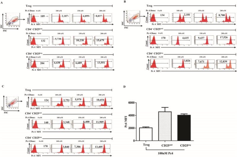
Treg cells from 3 subjects (defined as the highest 5% of CD4 cells expressing CD25) were sorted by flow cytometry and then incubated with Pc 4 (0–300 nM) in complete medium for 2 h. To measure monomeric Pc 4 levels, flow cytometric analysis was performed using a LSR II Flow Cytometer to obtain the mean fluorescence intensity (MFI) for each T cell subset. Pc 4 was excited by a broadband UV laser (335–365 nm) and fluorescence emission was collected with a 650-nm long-pass filter. Autofluorescence was subtracted from each sample. Three independent experiments are shown (A–C), indicating that CD4+ CD25high Treg cells incorporate less Pc 4 (lower Pc 4 MFI) than the proliferative populations of CD4+ CD25mid or CD4+ CD25neg T cells from the same subjects.